

By Peter G. Traber, M.D. on January 28, 2016
I wrote recently about why we rely on liver biopsies for the diagnosis of NASH and liver fibrosis and the need for alternative, non-invasive tests that enable us to diagnose and track the progression the disease. Today I’d like to explore the different non-invasive tests being developed. There are two basic approaches I will discuss: tests where blood (or serum) is tested for specific biomarkers, and the physical assessment of the liver using some form of imaging technology. I will use several prominent examples, but this will not cover every test being evaluated or available. I will also discuss tests of liver function and blood flow in future Perspectives.
Serum tests
Serum tests (serum is the clear fluid that results after blood clots) are already commonly used in support of liver biopsy for a variety of liver diseases. A liver biopsy can indicate that a patient has viral hepatitis, for example, but there is no definitive way of knowing whether it’s hepatitis A, B, C, D or E without a blood test.
Might we be able to take the next step and use a blood test without resorting to a biopsy? These tests are problematic from several fundamental standpoints. One reason is that you don’t necessarily know without rigorous empirical research whether what’s in the serum is reflective of what’s in the liver. For example, the galectin-3 protein is markedly increased in the liver tissues in animal models with NASH, but galectin-3 levels in the serum aren’t reflective of what’s going on in the tissue. The same holds true for many of the thousands of other proteins involved.
Despite this, there are many researchers who are trying to devise sophisticated blood tests that provide much of the information found on a liver biopsy, which has been called a “liquid biopsy.” Researchers have been using proteomic approaches to examine the thousands of different proteins and modified proteins that are in the blood to see whether there are tests that reflect either the etiology or the degree of damage in the liver. In other cases, researchers are measuring all the products of metabolism, called metabolomics, to investigate the correlation with biopsy findings. People are also looking at microRNA circulating in the blood, as well as micro vesicles, which are small pieces of cells that break off and circulate in the blood. These approaches, while promising, are far from well-defined enough to be used in clinical medicine.
To simplify our discussion, let’s concentrate on some of the currently available blood tests that look to determine the degree of fibrosis in the liver. Many approaches have been tried on this problem, such as testing for single compounds like hyaluronic acid or compiling multiple different serum tests together to determine a combined score, such as FibroTest® or the ELF (Enhanced Liver Fibrosis) test. Research shows that many of these tests correlate in various ways between serum levels and the degree of fibrosis, but they’re not very precise. They can tell you whether there’s little fibrosis or a lot of fibrosis, but they can’t tell you all the different gradations in between. There’s no solid evidence that they’re going to be useful in their current iterations for testing the effect of a drug, because such effects are likely to be found in those gradations.
We studied several of these serum tests in our Phase I study of GR-MD-02, and, as I discussed in an earlier CEO Perspective, we noticed that the alpha 2 macroglobulin protein, which is part of the FibroTest, appeared to be decreased with drug treatment. That said, in my view the accuracy, precision and reliability of serum tests have not gotten to the point where they could be used as a reliable surrogate for a biopsy in clinical trials of antifibrotic drugs.
Physical assessment of the liver
Another possibility for non-invasive testing of the liver is through physical assessment, often through some form of diagnostic imaging. A fibrotic liver has very different physical characteristics than a normal liver, and some of this can be assessed by external means.
There are some imaging techniques that wouldn’t work for the assessment of liver fibrosis. CT (computed tomography) and standard ultrasound tests show liver tissue, but they are really only effective for examining the overall structure of the liver, its size, and whether there are tumors — not that useful for determining fibrosis.
A variety of ultrasound-based tests are the best studied methods for staging liver fibrosis, including FibroScan®, acoustic radiation force impulse imaging (ARFI), and real-time shear wave elastography (SWE). All of these tests essentially evaluate the stiffness of the liver, because the more fibrosis a patient has, the stiffer the liver becomes. FibroScan, developed by Echosens and approved for diagnostic use in the US, measures the speed of a mechanical pulse as it goes through the liver, which correlates that to stiffness and produces a pressure measurement, kilopascals. While FibroScan only measures a core of liver tissue, it is about 100 times bigger than the area measured by a liver biopsy. Liver disease and fibrosis are often not uniform throughout the entire liver, so analyzing a small sample might lead to either over- or underestimating the amount of fibrosis. FibroScan has many desirable characteristics for a test of liver fibrosis in that it is quick, inexpensive, can be done in a provider office setting, and is painless. We utilized this test in the third cohort of our Phase 1 clinical trial (more here) and have included it in our two Phase 2 ongoing clinical trials, NASH-FX and NASH-CX.
Magnetic resonance elastography, or MRE, which is available from several manufacturers of imaging machines, uses low frequency vibrations applied to the patient. The MRI can pick up the perturbations as the pulse moves through the liver tissue. If the liver is very stiff, the pulse will move quickly. As with FibroScan, the results of MRE are reported in kilopascals and have been correlated to the degree of fibrosis as diagnosed through biopsy. It has the advantage of evaluating the entire liver, thereby eliminating sampling error of ultrasound based methods, and it simultaneously evaluates the liver for lesions such as tumors. While MRE compares favorably to FibroScan, and in some studies is more useful for staging fibrosis, it is much more expensive and less easily used in a clinical setting. We are also using this test in the NASH-FX study.
Software analyses of MRI data can be used to derive additional information. Such software-driven tests can measure various tissue parameters (so called multi-parametric tests) such as the amount of fluid, iron, fat and interstitial space – that is the space between the cells – in the liver. Since fibrosis and much of the inflammation are found in the interstitial space, this can be a useful test. Software combines all these elements to give a score that provides some indication of how much fibrosis there is in the liver. Similar to MRE, multi-parametric MRIs may be superior to a liver biopsy, because an MRI looks at the entire liver, slice by slice, while biopsy only samples 1/50,000th of the liver. We’re using multi-parametric MRI software called LiverMultiScan®, developed by Perspectum Diagnostics, as a monitoring tool in our NASH-FX trial.
There is good correlation between LiverMultiScan and the results of liver biopsies (1), and a recent paper shows that it also correlates with patient outcomes (2). In other words, you can predict which patients are going to have bad outcomes from their liver disease based on the LiverMultiScan results. Furthermore, the results of a LiverMultiScan change in response to treatment. The most dramatic change reported was in a patient who had NASH with fibrosis and then underwent an intestinal bypass as a weight loss procedure. The patient’s LiverMultiScan score and fibrosis went down dramatically with the loss of weight. While this test is approved for diagnostic use in Europe and the U.S., we still need a lot more data before it would be accepted as a regulatory end point by FDA for drug approval, but it is a promising approach towards non-invasive evaluation.
Each of these three imaging methods are approved by regulatory agencies for diagnostic use and are currently available to physicians, mostly in selected locations. However, they have not been used in prospective, therapeutic clinical trials to assess the efficacy of a drug in liver fibrosis. Well controlled clinical trials will be necessary, as we and other companies are currently undertaking, to validate these methods for use in drug approvals.
We’re not there yet, but I think we will see these sorts of approaches evolve quickly. The goal is to get to the point where we can characterize the underlying fibrosis even more quantitatively and effectively as with a liver biopsy. When we’re able to do that, it will eliminate the risk and the pain associated with a liver biopsy, and enable us to evaluate the whole liver, not just 1/50,000th of it, multiple times with a patient.
Reference List
1. Banerjee R, Pavlides M, Tunnicliffe EM, Piechnik SK, Sarania N, Philips R, et al. Multiparametric magnetic resonance for the non-invasive diagnosis of liver disease. J Hepatol 2014 Jan;60(1):69-77.
2. Pavlides M, Banerjee R, Sellwood J, Kelly CJ, Robson MD, Booth JC, et al. Multi-parametric magnetic resonance imaging predicts clinical outcomes in patients with chronic liver disease. J Hepatol 2015 Oct 12.
These “CEO Perspectives” are a regular feature of our communication activities and may contain forward looking statements within the meaning of the Private Securities Litigation Reform Act of 1995. These statements relate to future events and use words such as “may,” “might,” “could,” “expect” and others. These statements include those regarding the hope that Galectin Therapeutic’ s development program for GR-MD-02 will show that it can be both safe and effective when used in combination with other drugs for the treatment of patients with cancer. For a discussion of additional factors impacting Galectin’s business, see the Company’s Annual Report on Form 10-K for the year ended December 31, 2014, and subsequent filings with the SEC. You should not place undue reliance on forward-looking statements. Although subsequent events may cause its views to change, management disclaims any obligation to update forward-looking statements.
Make a Comment or Ask a Question
Related Articles
 2015 – 2016, Progress and Possibilities
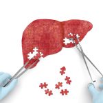 Results from the NASH-FX Study Underscore the Importance of Completing the NASH-CX Clinical Trial for Patients with NASH Cirrhosis
 Second Phase 2 clinical trial initiated for fatty liver disease with advanced fibrosis
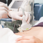 Why Non-Invasive Testing is Needed in Liver Fibrosis